Author BOB THOOLEN
Department of Veterinary Pathology, Branch Laboratory Animal Pathology and Special Animal Diseases, Veterinary Faculty, State University, 3508 TD Utrecht, The Netherlands.
Received for publication April 26, 1989 and in revised form July 19, 1989; accepted August 25, 1989 (9A1680).
In this study, BrdUrd labeling of S-phase cells in the small intestine and testes was accomplished using microwave irradiation. In this way crypt cells, spermatogonia, and Ley dig cells could be labeled using removable plastic-embedded sections and immunogold-silver staining (IGSS). By using short periods of microwave irradiation for incubation of the monoclonal antibodies and the protein A-colloidal gold solution, the detection of BrdUrd-labeled cells could be remarkably enhanced. A comparative study of BrdUrd labeled spermatogonia in the testis of a Cpb-N mouse that received both [3H]-thymidine and BrdUrd proved that 90% of the BrdUrd-labeled cells also showed [3H]-thymidine labeling. The radioactive [3H]-thymidine labeling was a time-consuming method of 4 weeks’ duration, whereas the BrdUrd labeled cells could be labeled, fixed, enhanced, and counterstained in less than 3 hr. This investigation proves that BrdUrd labeling of S-phase cells can be a reliable, reproductive, rapid, and non-radioactive alternative method for [3H]-thymidine labeling of proliferating cells. (] Histochem Cytochem 38:267-273, 1990)
Keywords
S-phase cells; Testes; BrdUrd; Spermatogonia; Leydig cells; lmmunogold-silver staining.
lntroduction
5-Bromodcoxyuridine (BrdUrd) incorporation into proliferating cells can be used to study the cell kinetics of different tissues. Several immunocytochemical studies in recent years detected BrdUrd in S-phase cells, used immunofluorescence techniques or immunoperoxidase staining to visualize the incorporated BrdUrd (Harms et al., 1987; Harms et al., 1986; Raza et al., 1985; Gratzner, 1982). The use of a microwave oven in this study enhanced the BrdUrd labeling of S-phase cells remarkably. When I used epipolarized light, the BrdUrd-labeled S-phase cells in the testes of mice could be visualized even more clearly. This report presents a quick, reliable, and specific labeling technique to demonstrate S-phase cells.
Materials and Methods
Testing of the BrdUrd Labeling Technique. Adult Cpb-N mice (15 weeks of age) weighing 30-35 g were used. They received an intraperitoneal injection of 5-BrdUrd (Sigma; St Louis, MO) at a dose of either 100 or 200 mg/kg body weight. The animals were sacrificed by cervical dislocation 2hr after injection. The small intestine and testes were removed and fixed in Carnoy’s fluid. The tissue was dehydrated, embedded in methyl methacrylate (K-plast medium), and cut at 2 µm. After removing the plastic with chloroform, hydrolization was performed in 1 N HCI at 60°C for 10 min and the sections washed in distilled water. The sections were neutralized with 0.1 N NaOH at 37°C for 1 min and rinsed during neutralization. After washing the sections in distilled water, the immunogold-silver staining procedure was used as follows.
- Rinse the sections in 0.15 M PBS (pH 7.4), 5 min.
- Block background staining with PBS with 0.4% BSA and 0.1% gelatin, 10 min.
- Rinse in 0.15 M PBS, 6 min.
- lncubate the slides with anti-BrdUrd monoclonal antibody diluted in PBS containing 0.1% BSA, 1:80. lrradiate (450 W) for 45 sec.
- Repeat steps described in 1, 2, and 3.
- lncubate with anti-mouse scrum diluted in PBS containing 0.1% BSA, 1:80. lrradiate (450 W) for 40 sec.
- Repeat step 3.
- lncubate with protein A-colloidal gold (9 nm) solution (PBS containing 0.1% BSA), 1:25. lrradiate (450 W) for 40 sec.
- Rinse 2 x 5 min, followed by distilled water for 10 min.
- Enhance the sections with Intense II kit at 18°C for 25 min.
- When enhancement is sufficient, wash the slides in distilled water for 10 min and counterstain with Gill’s hematoxylin no. 2 for 4 min.
- Rinse thoroughly in running tapwater for 5 min.
- Dehydrate in a graded series of ethanols to xylene.
- Mount the BrdUrd-labeled and colored tissue in Entellan.
The monoclonal antibody against BrdUrd (clone B44) mouse lgG1 heavy chain and kappa light chain was obtained from Becton-Dickinson (Mountain View, CA). Rabbit anti-mouse lgG serum (H + L) was obtained from Nordic (Tilburg, The Netherlands). Both Intense II kit and protein A-colloidal gold were received from Janssen Life Sciences (Beerse, Belgium). A microwave oven (Miele M 696) was used during the incubations of the monoclonal antibodies and the protein A-colloidal gold solution. The tissue slides were counterstained with Gill’s hematoxylin no. 2 (Polysciences; Warrington, PA).
Epipolarized light was used with the aid of a light microscope (Leitz orthoplan) to demonstrate the BrdUrd-labeled cells.
Comparison of BrdUrd and [3H]-Thymidine lncorporation. Two Cpb-N mice were injected IP with [3H]-thymidine (1.5 µCi/g body weight; Amersham, Poole, UK; sp. act. 55 Ci/mM) and BrdUrd (100 or 200 mg/kg body weight) simultaneously. Two hours later the animals were sacrificed by cervical dislocation and the testes were removed and fixed. Serial sections were made of the testes of the Cpb-N mice that received both BrdUrd and [3H]-thymidine. From two successive sections one was used for autoradiography while the other was processed according to the IGSS method described above.
The labeled spermatogonia were counted three times in each tubule, to be sure of the correct number of counts. Nine tubules were counted in both sections. The counts were made at a higher magnification (x 300), to be sure that the BrdUrd-labeled cells were the same as those showing [3H]-thymidine labeling. The tubules counted in both sections, BrdUrd-labeled and [3H]-thymidine-labeled sections, respectively, are marked with an asterisk (see Figure 4). The counts were made in sections of the Cpb-N mouse that received both BrdUrd (200 mg/kg body weight) and [3H]-thymidine (1.5 µCi/g body weight).
To compare the reliability of the BrdUrd- and [3H]-thymidine labeling, subsequent 2-µm sections were subjected to both labelings. One section was labeled according to the immunogold-silver staining procedure described above, to detect BrdUrd-labeled cells. The next section was covered with AR 10 stripping film (Kodak; Rochester, NY).
After 4 weeks of exposure the slides were developed in D19, fixed in a 24% (w/v) solution of sodium thiosulfate, and stained with hematoxylin. A Sony video camera was used in this experiment where counts were made of both BrdUrd- and [3H]-thymidine-labeled spermatogonia.
Results
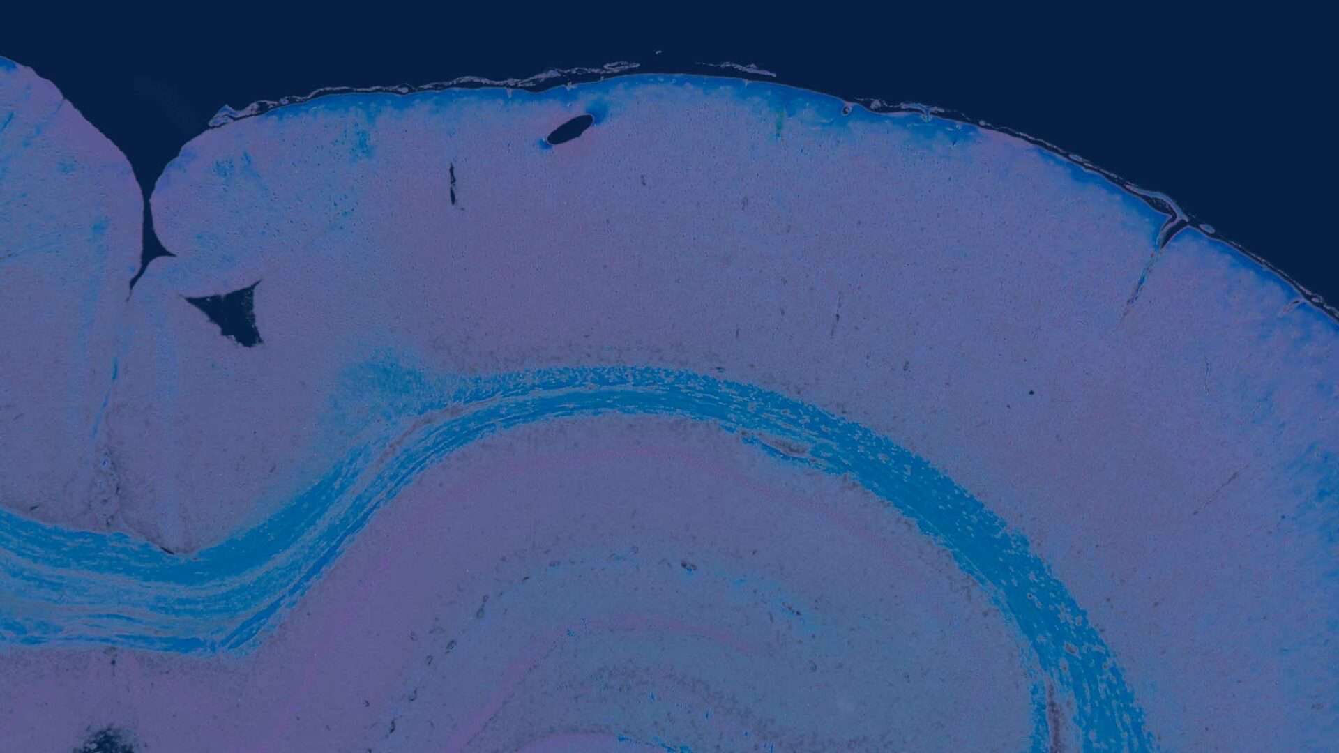
The results of this immunocytochemical study prove that BrdUrd labeling of spermatogonial stem cells and Leydig cells in the testes, and crypt cells of the small intestine of the Cpb-N mouse, is possible in plastic-embedded tissue sections. The results of the BrdUrd labeling technique are shown in Figures 1-5. The number of nuclei showing only [3H]-thymidine labeling was 162, while 166 nuclei showed only BrdUrd labeling. The number of nuclei showing both labels was 146. This indicates that 90% of the BrdUrd-labeled spermatogonia also showed [3H]-thymidine labeling (146/166 x 100%).


Discussion
In this investigative study, a comparison was made between [3H]-thymidine labeling and BrdUrd labeling of S-phase cells in the seminiferous tubules of the testes of Cpb-N mice. The results of this study are presented in Table 1. It is clear that the two labeling methods, BrdUrd and [3H]-thymidine labeling, are highly correlated (r = 0.99, p<0.001).
An innovation was the use of a microwave oven, which proved to be a very useful tool for enhancing the immunogold-silver staining. The microwave irradiations were carried out during the incubations of the monoclonal antibodies and the protein A-colloidal gold solution. Very short incubations of the anti-BrdUrd monoclonal antibodies and the anti-mouse serum could be performed (45 and 40 sec, respectively). The incubation of the protein A-colloidal gold solution was also of very short duration (40 sec). When I used shorter incubation times, no specific labeling of S-phase cells could be seen. Longer incubation times for both monoclonal antibodies and protein A-colloidal gold solution gave a diffuse brown background staining on the slides. The slight background staining seen in Figures 1, 2B, 3B, and 5 is the result of the instability of the Intense II kit, which is temperature sensitive. Nevertheless, by following the procedures detailed in Materials and Methods,
Table 1. Counts of spermatogonia labeled with BrdUrd, [3H]-thymidine, or both (see Figures 4A and 4B)
| Tubule no. | BrdUrda | [3H]-Thymidineb | BrdUrd and [3H]-thymidinec |
| 1 | 0 | 0 | 0 |
| 2 | 5 | 3 | 3 |
| 3 | 52 | 48 | 47 |
| 4 | 22 | 22 | 19 |
| 5 | 0 | 0 | 0 |
| 6 | 15 | 15 | 13 |
| 7 | 2 | 2 | 2 |
| 8 | 15 | 14 | 13 |
| 9 | 55 | 58 | 49 |
a Counts of BrdUrd-labeled spermatogonia.
b Counts of [3H]-thymidine-labeled spermatogonia.
c Counts of spermatogonia labeled with both BrdUrd and [3H]-thymidine. Correlation coefficient of both labeling methods: r = 0.99 (p<0.001).
specific BrdUrd labeling of S-phase cells in the mouse testes could be obtained. I repeated the BrdUrd labeling experiment several times, and when the IGSS procedure was followed correctly all experiments were successful. The use of plastic-embedded sections has the advantage that the plastic can easily be removed with chloroform, thus making the antigens became accessible to the antibodies used in this BrdUrd labeling procedure.
Sometimes more spermatogonia showed BrdUrd labeling than [3H]-thymidine labeling when the Cpb-N mouse had received IP injections of both BrdUrd and [3H]-thymidine (see Table 1). An explanation of why more nuclei was labeled with BrdUrd cannot be given at this time. On the basis of this finding, one may say that BrdUrd labeling is a non-radioactive, reliable, and much less time-consuming labeling technique for detecting S-phase cells in the testes of laboratory mice. Little is known about the mutagenicity or the carcinogenicity of BrdUrd, although some authors believe that BrdUrd has both mutagenic and carcinogenic properties (Napalkov et al., 1989; deFazio et al., 1988). Nevertheless, others have proved that BrdUrd labeling can be a reliable and very useful tool in cell kinetics studies (Kikuyama et al., 1988; Riccardi et al., 1988; Silvestrini et al., 1988).
The possible applications of the BrdUrd labeling technique are numerous. It has been used for cell kinetics studies in various tissues (Harms et al., 1987; Jonkers and Dongen, 1986; Raza et al., 1985; Gratzner, 1982) and for clinical studies of human carcinogenesis (Kyung et al., 1986; Lokhorst et al., 1986), and in the future may prove to be very useful in both human and animal pathological studies.
Acknowledgments
I wish to thank Mr A.N. van Rijn for making the photographs, and Prof P. Zwart and Theo Bakker for reviewing the manuscript.
Literature Cited
Cho KG, Hoshino T, Nagashima T, Murovic JA, Wilson CB (1986): Prediction of tumor doubling time in recurrent meningiomas. J Neurosurg 65:790. https://doi.org/10.3171/jns.1986.65.6.0790
de Fazio A, Musgrove EA, Tattersall MH (1988): Flow cytometric enumeration of drug-resistant tumor cells. Cancer Res 48:6037
Gratzner HG (1982): Monoclonal antibody to 5-bromo and 5-iododeoxyuridine: a new reagent for detection of DNA replication. Science 218:474 DOI: 10.1126/science.7123245
Harms G, van Goor H, Koudstaal J, de Ley L, Hardon KMJ (1987): Simultaneous immunohistochemical demonstration of antigen expression and 5-bromodeoxyuridine incorporation in plastic embedded sections. Histochemistry 86:393
Harms G, van Goor H, Koudstaal J, de Ley L, Hardonk MJ (1986): Immunohistochemical demonstration of DNA-incorporated 5-bromodeoxyuridine in frozen and plastic embedded sections. Histochemistry 85:139
Jonkers B, Dongen PAM (1986): Antilichamen als diagnostisch gereedschap. Beerse, Belgium, Janssen Medisch-Wetenschappelijk Nieuws, 13
Kikuyama S, Kubota T, Watanabe M, lshibiki K, Abe O (1988): Cell kinetic study of human carcinomas using bromodeoxyuridine. Cell Tissue Kinet 21:15. https://doi.org/10.1111/j.1365-2184.1988.tb00767.x
Lokhorst HM, Boom SE, Bast BJEG, Ballieux RE (1986): Determination of the plasma cell labelling index with bromodeoxyuridine in a double fluorescence technique. Br J Haematol 64:271. https://doi.org/10.1111/j.1365-2141.1986.tb04119.x
Napalkov NP, Anisimov VN, Likhachev AJ, Tomatis L (1989): 5- Bromodeoxyuridine-induced carcinogenesis and its modification by persistent estrus syndrome, unilateral nephrectomy, and X-radiation in rats. Cancer Res 49:318. http://cancerres.aacrjournals.org/content/49/2/318
Raza A, Ucar K, Bhayana R, Kempski M, Preisler A (1985): Utility and sensitivity of anti-BrdU antibodies in assessing S-phase cells compared to autoradiography. Cell Biochem Funct 3:149. https://doi.org/10.1002/cbf.290030212
Riccardi A, Danova M, Wilson G, Ucci G, Dormer P, Mazzini G, Brugnatelli S, Girino M, McNally NJ, Ascari E (1988): Cell kinetics in human malignancies studied with in vivo administration of bromodeoxyuridine and flow cytometry. Cancer Res 48:6238
Silvestrini R, Costa A, Veroni S, Del Bino G, Persici P (1988): Comparative analysis of different approaches to investigate cell kinetics. Cell Tissue Kinet 21:123. https://doi.org/10.1111/j.1365-2184.1988.tb00778.x