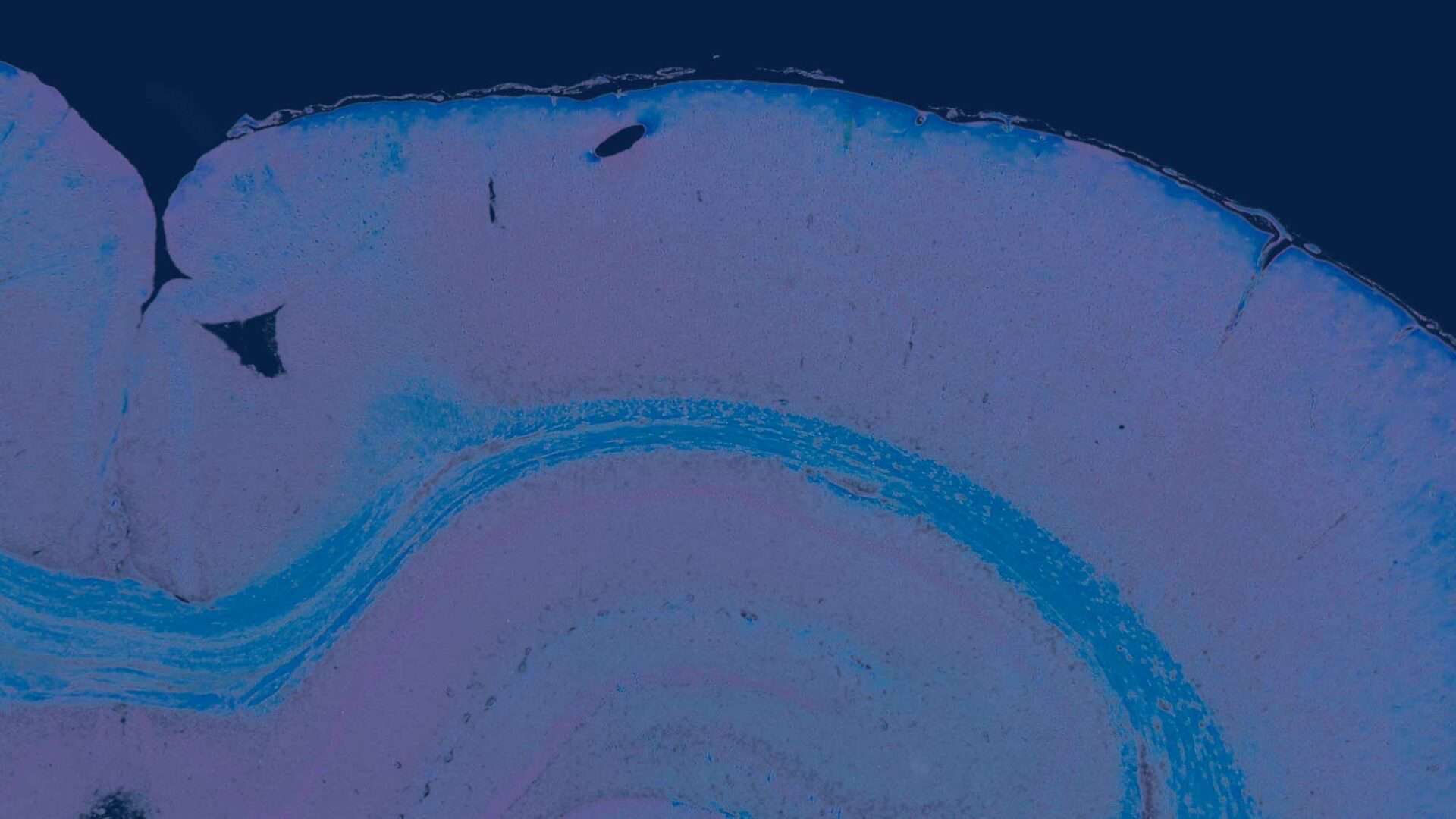
Digital Pathology, also referred to as Telepathology and Whole Slide Imaging (W.S.I.), is the process of producing high resolution digital images from tissue sections on glass slides, and subsequent evaluation. These glass slides are normally examined under a microscope by a pathologist as part of the diagnostic process. The emergence of digital pathology now means that digital images are stored on secure servers and can be viewed on computer monitors; enabling pathologists to work remotely and to collaborate with other colleagues and/or when second opinions are needed.
Conventional Histopathology
Histopathology is conventionally performed by certified pathologists using a light microscope. This histopathological examination is not an exercise in “picture matching” for it represents a careful step by step evaluation of tissue and cellular patterns. This includes assessment of size, shape, staining characteristics and topographical tissue and cell organizations and integration into meaningful biological conclusions, with considerations of the overall macroscopic and microscopic pathology as well as other related medical disciplines. Species specificity, anatomy, artefacts, control references and experience of the study pathologist are considered key factors in this process. Additional semi-quantitative techniques and artificial intelligence (A.I.) can also be valuable additional techniques for research purposes and decision support.
Digital Pathology: the future of diagnostics
We have been following the developments of Digital Pathology over the recent years, and it seemed good for peer review and/or educational purpose (e.g. Academic Hospitals, large CROs) of limited number of slides, and long-distance tele-communication. In the past, next to conventional light microscopy, also peer review was done faster in a conventional way by reviewing the glass slides. In addition, in the past there were sometimes problems with proper scanning of every slide which was time consuming.Nowadays, however, the quality of images generated by using Digital Pathology is as good as conventional review of slides for all organ systems using digital pathology. The UMCU Academic hospital in Utrecht, The Netherlands was one of the the first hospitals world-wide going fully digital in pathology with a future-proof complete digital archive (2015).During and after this time, scan speed, resolution and quality of scans from glass slides (W.S.I.) have been markedly improved. In recent years, substantial increased amount of W.S.I. validation literature is published on this subject. For both in clinical pathology (human) and pre-clinical veterinary pathology practices, things have improved significantly with demonstration of excellent diagnostic concordance between W.S.I. and conventional microscopy. Nowadays, the application of Digital Pathology, when well performed, can match equally with conventional light microscopy in toxicologic pathology for the safety evaluation in toxicology studies in the development of new vaccines, medicines and chemicals.
Digital Pathology at Global Pathology Support
As part of GPS continuing commitment to enable our clients to reach their development goals, we are now excited, next to conventional histopathology using the light microscope, to also deliver innovative GLP validated Digital Pathology; Full Histopathology Evaluations and Peer Review solutions from preclinical toxicology studies on a Global scale. This option (Digital Pathology) can now also be performed from scans of histology slides from a large number of whole slide image formats (e.g. Hamamatsu, 3DHistech, Olympus, Aperio/Leica, Ventana/Roche, Philips, Zeiss scanners etc. ).For more information: bob.thoolen@gpstoxpath. +31 (0)70 3142404
Bibliography
- Abels E, Pantanowitz L, Aeffner F, et al. Computational pathology definitions, best practices, and recommendations for regulatory guidance: a white paper from the Digital Pathology Association. The Journal of Pathology 2019;249(3):286-294.
- Aeffner F, Sing T, Turner OC. Special Issue on Digital Pathology, Tissue Image Analysis, Artificial Intelligence, and Machine Learning: Approximation of the Effect of Novel Technologies on Toxicologic Pathology. Toxicol Pathol 2021;49(4):705-708.
- Al-Janabi S, Horstman A, van Slooten H-J, et al. Validity of whole slide images for scoring HER2 chromogenic in situ hybridisation in breast cancer. J Clin Pathol 2016;69(11):992. (
- Al-Janabi S, Huisman A, Jonges GN, et al. Whole slide images for primary diagnostics of urinary system pathology: a feasibility study. J Ren Inj Prev 2014;3(4):91-96.
- Araújo ALD, do Amaral-Silva GK, Pérez-de-Oliveira ME, et al. Fully digital pathology laboratory routine and remote reporting of oral and maxillofacial diagnosis during the COVID-19 pandemic: a validation study. Virchows Archiv : an international journal of pathology 2021;479(3):585-595.
- Azam AS, Miligy IM, Kimani PK-U, et al. Diagnostic concordance and discordance in digital pathology: a systematic review and meta-analysis. J Clin Pathol 2021;74(7):448-455.
- Baidoshvili A, Stathonikos N., Freling G, et al. Validation of a whole-slide image-based teleconsultation network. Histopathology 2018;73(5):777-783.
- Baidoshvili A, Bucur A, van Leeuwen J, et al. Evaluating the benefits of digital pathology implementation: time savings in laboratory logistics. Histopathology 2018;73(5):784-794.
- Bejnordi BE, Balkenhol M, Litjens G, et al. Automated Detection of DCIS in Whole-Slide H&E Stained Breast Histopathology Images. IEEE Trans Med Imaging 2016;35(9):2141-2150.
- Bejnordi BE, Karssemeijer N, Timofeeva N, et al. Quantitative analysis of stain variability in histology slides and an algorithm for standardization. Prog. Biomed. Opt. Imaging – Proc. SPIE 2014;9041:7.
- Bejnordi BE, Litjens G, Hermsen M, et al. A multi-scale superpixel classification approach to the detection of regions of interest in whole slide histopathology images. Proc. SPIE, Med Imag 2015:6.
- Bertram CA, Firsching T, Klopfleisch R. Virtual microscopy in histopathology training: Changing student attitudes in 3 successive academic years. Journal of Veterinary Medical Education 2018;45(2):241-249.
- Bertram CA, Stathonikos N, Donovan TA, et al. Validation of digital microscopy: Review of validation methods and sources of bias. Vet Pathol 2022;59(1):26-38.
- Betmouni S. Diagnostic digital pathology implementation: Learning from the digital health experience. Digit Health 2021;7.
- Bongaerts O, Clevers C, Debets M, et al. Conventional Microscopical versus Digital Whole-Slide Imaging-Based Diagnosis of Thin-Layer Cervical Specimens: A Validation Study. J Pathol Inform 2018;9:29.
- Borowsky AD, Bishop JW, Darrow MA, et al. Digital whole slide imaging compared with light microscopy for primary diagnosis in surgical pathology: A multicenter, double-blinded, randomized study of 2045 cases. Arch Pathol Lab Med 2020;144(10):1245-1253.
- Caputo A, D’Antonio A. Digital pathology: the future is now. Ind J Pathol Microbiol 2021;64(1):6-7.
- Cimadamore A, Scarpelli M, Montironi R, et al. Digital pathology and COVID-19 and future crises: pathologists can safely diagnose cases from home using a consumer monitor and a mini PC. J Clin Pathol 2020;73(11):695-696.
- Clarke E, Doherty D, Randell R, et al. Faster than light (microscopy): superiority of digital pathology over microscopy for assessment of immunohistochemistry. J Clin Pathol 2022;0:1-6.
- Evans AJ, Brown RW, Bui MM, et al. Validating Whole Slide Imaging Systems for Diagnostic Purposes in Pathology. Arch Pathol Lab Med 2022;146(4):440-450.
- FDA. The US Food and Drug Administration temporarily relaxes regulations around digital pathology to facilitate remote reviewing of digital pathology slides during the COVID-19 pandemic. globenewswire 2020.
- Forest T, Aeffner F, Bangari DS, et al. Scientific and Regulatory Policy Committee Brief Communication: 2019 Survey on Use of Digital Histopathology Systems in Nonclinical Toxicology Studies. Toxicol Pathol 2022;50(3):397-401.
- Geijs DJ, Intezar M, van der Laak JAWM, et al. Medical Imaging: Digital Pathology – Automatic color unmixing of IHC stained whole slide images. Med Imag 2018: Dig Pathol 2018:165-171.
- Ghezloo F, Pin-Chieh W, Kerr KF, et al. An analysis of pathologists viewing processes as they diagnose whole slide digital images. J Pathol Inf 2022;13:1-6.
- Go H. Digital Pathology and Artificial Intelligence Applications in Pathology. Brain Tumor Res Treat 2022;10(2):76-82.
- Hanna MG, Ardon O, Reuter VE, et al. Integrating digital pathology into clinical practice. Mod Pathol 2021;35(2):152-164.
- Hanna MG, Reuter VE, Ardon O, et al. Validation of a digital pathology system including remote review during the COVID-19 pandemic. Mod Pathol 2020;33(11):2115-2127.
- Jacobsen M, Lewis A, Baily J, et al. Utilizing Whole Slide Images for the Primary Evaluation and Peer Review of a GLP-Compliant Rodent Toxicology Study. Toxicol Pathol 2021;49(6):1164-1173.
- Jahn SW, Plass M, Moinfar F, et al. Digital pathology: Advantages, limitations and emerging perspectives. J Clin Med 2020;9(11):1-17.
- Jones-Hall YL, Skelton JM, Adams LG. Implementing Digital Pathology into Veterinary Academics and Research. J Vet Med Educ 2021;49(5):547-555.
- Kim YJ, Roh EH, Park S. A literature review of quality, costs, process-associated with digital pathology. J Exerc Rehabil 2021;17(1):11-14.
- Klaver E, van der Wel M, Duits L, et al. Performance of gastrointestinal pathologists within a national digital review panel for Barrett’s oesophagus in the Netherlands: results of 80 prospective biopsy reviews. J Clin Pathol 2021;74(1):48-52.
- Korzynska A, Roszkowiak L, Zak J, et al. A review of current systems for annotation of cell and tissue images in digital pathology. Biocybern Biomed Eng. 2021;41(4):1436-1453.
- Kumar N, Gupta R, Gupta S. Whole Slide Imaging (WSI) in Pathology: Current Perspectives and Future Directions. J Digit Imaging 2020;33(4):1034-1040.
- Laurent-Bellue A, Redon MJ, Posseme K, et al. Four-Year Experience of Digital Slide Telepathology for Intraoperative Frozen Section Consultations in a Two-Site French Academic Department of Pathology. Am J Clin Pathol 2020;154(3):414-423.
- Limperio V, Brambilla V, Cazzaniga G, et al. Digital pathology for the routine diagnosis of renal diseases: a standard model. J Nephrol 2021;34(3):681-688.
- Parwani AV. Next generation diagnostic pathology: use of digital pathology and artificial intelligence tools to augment a pathological diagnosis. Diagn Pathol 2019;14(1):138.
- Post RS, van der Laak JAWM, van der Sturm B, et al. The evaluation of colon biopsies using virtual microscopy is reliable. Histopathology 2013;63:114.
- Rajaganesan S, Kumar R, Rao V, et al. Comparative Assessment of Digital Pathology Systems for Primary Diagnosis. J Pathol Inf 2021;12(1):1-15.
- Retamero JA, Aneiros-Fernandez, J., del Moral, R. G. . Complete Digital Pathology for Routine Histopathology Diagnosis in a Multicenter Hospital Network. Arch Pathol Lab Med 2020;144(2):221-228.
- Retamero JA, Aneiros-Fernandez J, Del Moral RG. Microscope? No, thanks: User experience with complete digital pathology for routine diagnosis. Arch Pathol Lab Med 2020;144(6):672-673.
- Schumacher VL, Aeffner F, Barale-Thomas E, et al. The Application, Challenges, and Advancement Toward Regulatory Acceptance of Digital Toxicologic Pathology: Results of the 7th ESTP International Expert Workshop (September 20-21, 2019). Toxicol Pathol 2021;49(4):720-737.
- Stathonikos N, Veta M, Huisman A, et al. Going fully digital: Perspective of a Dutch academic pathology lab. Journal of pathology informatics 2013;4(1):15.
- Stathonikos N, Nguyen TQ, Spoto CP, et al. Being fully digital: perspective of a Dutch academic pathology laboratory. Histopathology 2019;75(5):621-635.
- Stathonikos N, ,, Van Varsseveld NC, Vink A, et al. Digital pathology in the time of corona. J Clin Pathol 2020;73(11):706-712.
- Unternaehrer J, Zlobec I, Grobholz R, et al. Current opinion, status and future development of digital pathology in Switzerland. J Clin Pathol 2020;73(6):341-346.
- Veta M, van Diest PJ, Willems SM, et al. Assessment of algorithms for mitosis detection in breast cancer histopathology images. Medical Image Analysis 2015;20(1):237-248.
- Vodovnik A, Riste T, Sund BS. Digital Pathology During a Pandemic. J Pathol Inform 2020;11:1-2.
- Williams B, Hanby A, MillicanSlater R, et al. Digital pathology for primary diagnosis of screendetected breast lesions – experimental data, validation and experience from four centres. Histopathology 2020;76(7):968-975.
- Williams BJ, Treanor D. Practical guide to training and validation for primary diagnosis with digital pathology. J Clin Pathol 2020;73(7):418-422.
- Zarella MD, Bowman D, Aeffner F, et al. A Practical Guide to Whole Slide Imaging: A White Paper From the Digital Pathology Association. Arch Pathol Lab Med 2019;143(2):222-234.
